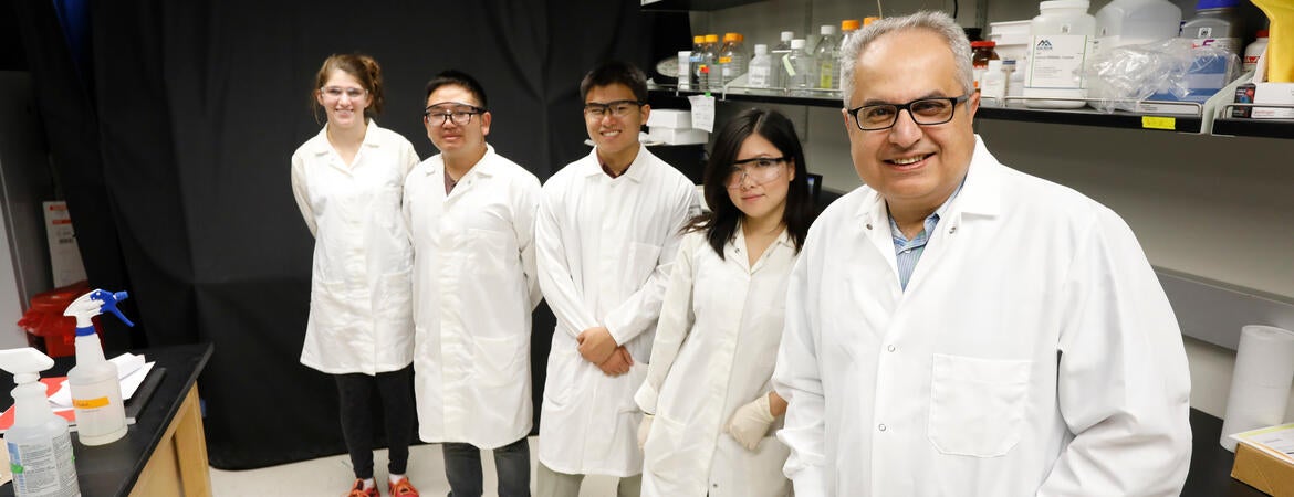
UCR Bioengineering Professor Bahman Anvari and University of Texas MD Anderson Cancer Center School of Medicine Professor Vikas Kundra received a $300,000 grant from the NSF to investigate the transformative potential of dual imaging modality using magnetic resonance (MR) and fluorescence for early detection, staging, and guided-resection of ovarian tumor implants.
The dual imaging modality method used is based on an innovative liposomal nanoparticle system that contains gadolinium (Gd), and a new brominated cyanine dye (BrCy106) as the respective MR and optical contrast agents. The goal of the researchers is to develop both early detection and localization of small ovarian tumors prior to surgery by MR imaging (MRI), followed by near infrared flourescience imaging (NIRDFI) at surgery to aide staging and guide surgical removal of all tumors which cannot otherwise be visualized.
The research team will optimize the formulation of the nanoparticle system for maximum relaxivity as well as fluorescence quantum yield by experimenting with various concentrations of Gd and BrCy106. The feasibility of the optimized nanoparticles for MRI and NIRFI will be evaluated with mice with ovarian cancer cell implantations to mimic early stage tumors and late stage disease.